The meniscus is a wedge-shaped piece of cartilage situated within the knee joint, positioned between the femur (thighbone) and the tibia (shinbone). There are two menisci in each knee: the medial meniscus, located on the inner side of the tibia, and the lateral meniscus, found on the outer side. The medial meniscus resembles the letter "C" when viewed from the top, while the lateral meniscus has a shape resembling the letter "D." Both menisci are firmly attached to the top surface of the tibia, with the peripheral portions connected to the knee joint capsule. Functionally, the menisci serve as vital shock absorbers for the knee joint. They help to distribute compression forces from the femur over a wider area on the tibia, thereby reducing the impact on the joint during movement. Additionally, the menisci contribute to knee stability by aiding in the smooth movement of the joint surfaces. The medial meniscus bears approximately 50% of the load applied to the medial compartment of the knee, while the lateral meniscus absorbs up to 80% of the load on the lateral compartment. Throughout various activities such as walking, running, or jumping, forces exerted on the knee can vary significantly. For instance, during walking, forces on the knee may increase to 2-4 times the body weight, while running may result in forces up to 6-8 times the body weight. The menisci play a crucial role in managing these forces and maintaining the integrity and function of the knee joint.
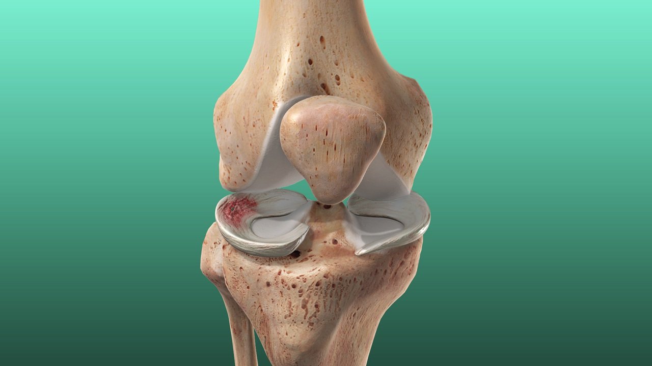
Each meniscus comprises three regions: the anterior (front), posterior (back), and middle regions. The anterior region includes the anterior root, anterior horn, and anterior part of the body, while the posterior region contains the posterior root, posterior horn, and posterior part of the body. The middle region forms the body of the meniscus. The roots firmly attach the front and back ends of both menisci to the top of the tibia on the inner (medial) and outer (lateral) sides. The entire periphery of the medial meniscus is attached to the joint capsule and a portion of the inner ligament (deep medial collateral ligament). The lateral meniscus's peripheral portion is also attached to the knee joint capsule, except for a small area where the popliteus tendon separates it from the capsule. This difference in peripheral attachment makes the lateral meniscus relatively more mobile than the medial meniscus. The menisci receive their blood supply from peripheral blood vessels that travel inward from the periphery to the central free portion. This unique pattern of blood supply creates three zones of variable blood supply within the meniscus: the outer or peripheral zone, which has the maximum blood supply and is hence called "red on red"; the middle third zone, which has fewer blood vessels compared to the peripheral portion and is termed "red on white"; and the inner free edge of the meniscus, which lacks blood supply and is thus referred to as the "white on white" zone.
Meniscus tears are frequently observed knee injuries among athletes and individuals engaged in active pursuits, particularly contact sports. These tears typically result from a sudden bending or twisting motion of the knee, leading to trauma. Such traumatic tears can affect either the medial or lateral meniscus, or sometimes both simultaneously. In elderly individuals, degenerative meniscal tears are more common due to the natural weakening of the meniscus with age. These tears often manifest in the posterior horn of the medial meniscus and are frequently complex in nature.
A torn meniscus can result in various symptoms, including pain, swelling, and stiffness in the knee. Patients may experience a sensation of catching or locking in the joint, hindering their ability to move the knee through its full range of motion. Some individuals may develop a limp or encounter difficulty when walking. Meniscal tears can occur independently following an injury, or they may coincide with other knee injuries such as ligament tears, cartilage damage, or fractures in the vicinity of the knee.
Meniscal tears are typically diagnosed through a thorough medical history, clinical examination, X-rays, and a knee MRI scan. In some cases, they may also be detected during knee arthroscopy. X-rays may appear normal in acute cases or reveal fractures, while they may also indicate calcium deposits in the meniscus in conditions like pseudo-gout (chondrocalcinosis). MRI scans are crucial for confirming the presence and type of meniscal tear, identifying any additional injuries, and detecting peri-meniscal cysts. Meniscal tears observed on knee MRI scans are categorized into three grades. Grades I and II are generally less severe and may not be evident during arthroscopic examination. Grade III denotes a significant meniscus tear, which arthroscopy can accurately diagnose close to 100 percent of the time.
Meniscal tears come in various types, characterized by their appearance and location within the knee joint. These include longitudinal tears, bucket handle tears (where the torn portion flips inward), radial tears, parrot beak tears, horizontal tears, oblique or flap tears, root tears, and complex tears (a combination of other tear patterns). Additionally, there are degenerative tears, which occur in older individuals with pre-existing cartilage degeneration, presenting as ragged tears.
Treatment for meniscal tears can be either non-surgical or surgical, depending on factors such as the type, size, and location of the tear, as well as the patient's age and activity level. Initially, patients are prescribed suitable painkillers and anti-inflammatory medications to alleviate pain and swelling. They are advised to rest the knee and apply ice packs regularly. Physiotherapy exercises are taught to patients, and they are encouraged to resume knee movements as tolerated. Some patients may require crutches for walking. Non-surgical treatment is typically recommended for those with small tears, peripheral tears, Grade I or II tears on MRI, and degenerative meniscal tears in the elderly. With this approach, pain and swelling typically subside over 4 to 6 weeks, and patients can return to their normal activities within 2 to 3 months
Surgery for meniscal tears is typically recommended in specific cases, such as Grade III tear patterns on MRI, displaced bucket handle tears, tears involving the anterior or posterior roots of the meniscus, tears associated with discoid menisci, and those linked with fractures or other ligament injuries. Additionally, surgical intervention may be advised for patients whose symptoms persist despite initial conservative treatments, like degenerative posterior horn tears in older individuals. Surgical options include partial meniscectomy, where the damaged portion of the meniscus is removed, often used for complex tears or failed repairs. Meniscus repair involves suturing torn tissue, suitable for certain tear patterns, while meniscus replacement or transplantation inserts complete meniscal tissue harvested from cadavers, typically for younger patients with previous meniscectomies and minimal arthritis. Each approach aims to address the tear's severity and location, promoting knee function and reducing long-term complications like degenerative arthritis.
Articular cartilage is a specialized tough connective tissue that covers the ends of bones within joints. Comprising chondrocyte cells embedded in a matrix of collagen fibers and proteins, it facilitates smooth movement between bones with minimal friction. Unlike many tissues, articular cartilage lacks blood or nerve supply, appearing white in color due to this absence. It receives nutrients through diffusion and is maintained and repaired by its contained cells. High fluid content contributes to its self-lubricating nature, offering the lowest coefficient of friction among natural and man-made materials. However, due to its lack of blood supply, injuries to articular cartilage cannot heal on their own, and pain may not be felt until lesions become deep enough to affect the underlying bone, which does have nerve supply.
Injuries or lesions of the articular cartilage can arise from various causes, including trauma such as blunt impacts, traumatic patellar dislocation, or polytraumatic injuries. Additionally, axial malalignment of the knee can result in overloading of specific areas, while fractures of the articular surfaces of the femur, tibia, or patella can directly damage the cartilage. Surgical procedures like partial or total meniscectomy may increase stress in affected compartments. Instability due to injuries to ligaments like the ACL or PCL can accelerate wear and tear, especially at a younger age. Conditions like osteochondritis dissecans, osteoarthritis, rheumatoid arthritis, and genetic factors can also contribute to cartilage damage. Furthermore, factors such as obesity, cartilage tumors, and repeated micro-trauma can exacerbate joint deterioration and increase the risk of developing arthritis.
Outerbridge's classification system, based on arthroscopy findings, provides a straightforward and clinically relevant method for assessing articular cartilage lesions. This system aids in understanding the severity of the lesion and guides treatment planning. According to Outerbridge's Classification of Chondral Lesions, Grade 0 represents normal articular cartilage, while Grade I indicates softening, blistering, or swelling of the cartilage. Grade II involves partial thickness defects with fissures and clefts not reaching the subchondral bone and typically less than 1.5 cm in diameter. Grade III denotes full-thickness fissures that extend to the subchondral bone, typically larger than 1.5 cm. Grade IV indicates exposed subchondral bone.
Articular cartilage injuries are most frequently observed in the knee joint, although other joints like the elbow, wrist, ankle, shoulder, and hip can also be affected. In acute cases, patients may experience pain, swelling, difficulty walking, and reduced mobility in the knee joint. They may also report sensations of knee locking or giving way, accompanied by limping while walking. Physical examination may reveal a swollen and tender knee, with bruising around the joint and limited painful movements. Signs of co-existing injuries, such as meniscal or ligament tears, may also be present. In the chronic stage, patients may continue to experience pain, intermittent swelling, stiffness, and a sense of instability or clicking in the knee. Episodes of knee locking may occur, particularly during activities such as stair climbing, squatting, kneeling, or playing sports, which often exacerbate the pain.
Articular cartilage injuries are diagnosed through a comprehensive process involving detailed medical history, thorough clinical examination, and appropriate investigations such as knee X-rays and MRI scans. While X-rays cannot visualize articular cartilage due to its lack of calcium, they provide valuable information on limb alignment, presence of loose bodies, fractures, joint space reduction, meniscal cartilage calcification, osteophyte formation, patellar tilting or mal-tracking, bone defects at the joint level, and cystic changes within subchondral bones. Modern MRI scans offer a detailed view of articular cartilage, detecting even minor lesions. Various MRI scoring techniques focus on assessing lesion size, location, and associated abnormalities in subchondral, cartilaginous, bony, and meniscal structures. MRI imaging highlights pathologies such as cartilage defects, injuries to the subchondral plate, bony lesions like osteochondritis dissecans or fractures, and secondary damages such as ligament tears. MRI plays a crucial role in planning the definitive treatment for patients with articular cartilage injuries.
Treatment options for articular cartilage injuries are diverse and aim to facilitate the healing and restoration of the affected area while enhancing joint function and minimizing the risk of osteoarthritis development. Surgical interventions such as arthroscopic debridement involve smoothing damaged cartilage to alleviate irritation, while marrow stimulation techniques like micro-fractures promote the growth of fibrocartilage to fill the defect. Osteochondral transfer procedures, including autologous techniques like OATS, replace the damaged cartilage with healthy tissue harvested from non-weight-bearing areas. Autologous chondrocyte implantation (ACI) and matrix-guided techniques like MACI offer biological regeneration by implanting cultured chondrocytes within scaffolds to promote precise cartilage regrowth. Alternatively, transplantation of fresh osteochondral allografts from cadaver donors provides larger grafts for extensive defects. These interventions aim to restore cartilage integrity, alleviate symptoms, and improve joint function, thereby enhancing the patient's quality of life.
Address
8/92, Sector 8, Ismailganj, Indira Nagar, Lucknow, Uttar Pradesh 226016
Monday to Friday
10am - 8pm
Appoinments
+91 - 8840223370